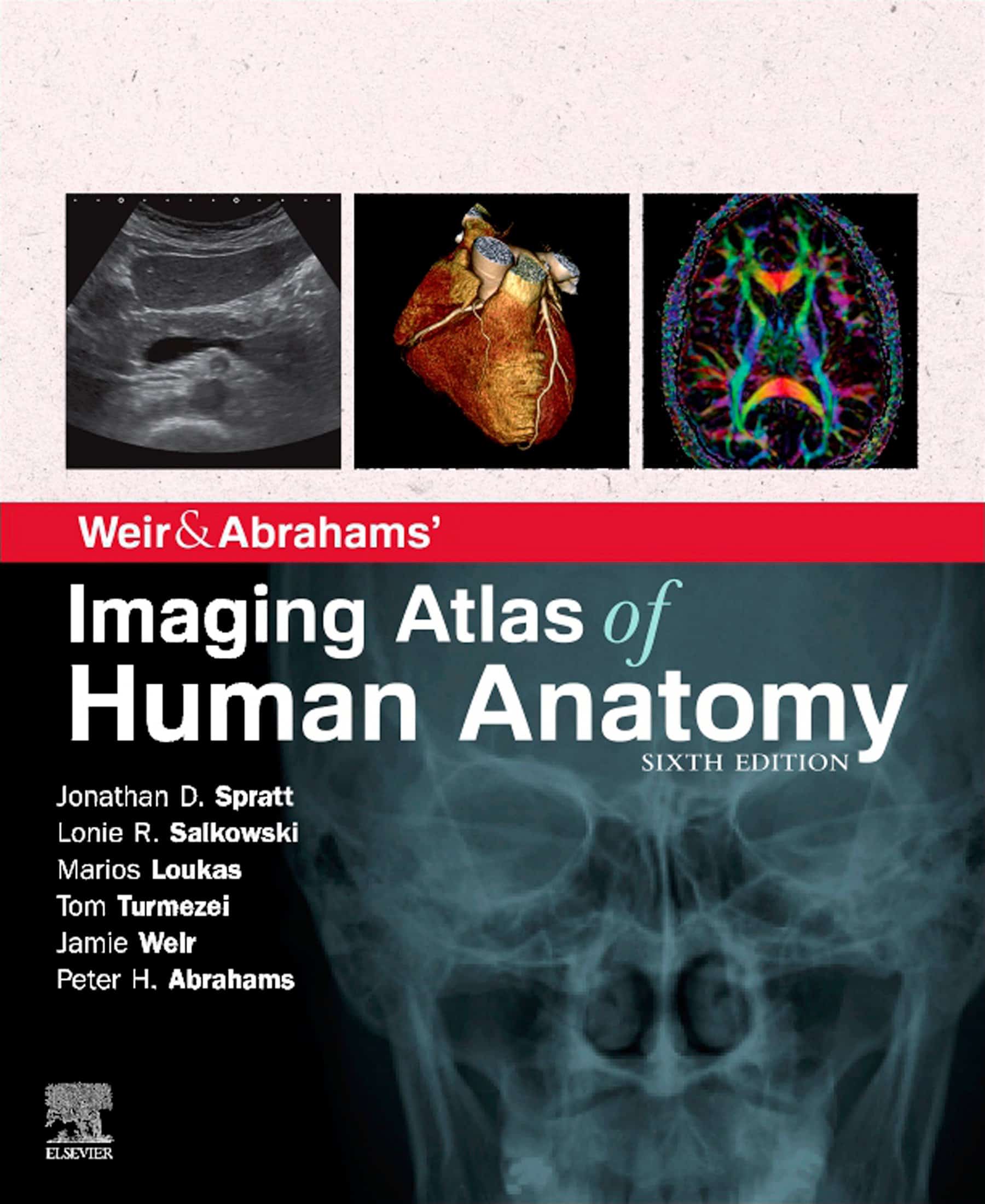
Imaging Atlas of Human Anatomy, 6th Edition, (PDF) is the best up-to-date imaging information for a complete and three-dimensional understanding of utilized human anatomy Imaging is ever extra important to anatomy training and all through trendy drugs. Building on the success of prior editions, this utterly revised sixth version offers an impressive basis for understanding utilized human anatomy, offering a whole view of the buildings and relationships inside the entire physique, utilizing very trendy imaging strategies. All associated imaging modalities are included, from plain radiographs to extra superior imaging of ultrasound, CT, purposeful imaging, MRI, and angiography. Coverage is additional improved by a rigorously chosen vary of BONUS digital content material, together with scientific circumstances and pictures, ultrasound movies, cross-sectional imaging stacks, labeled radiograph ‘slidelines’ and take a look at-your self supplies. Uniquely, key syllabus picture units at the moment are underlined all through to help environment friendly research, along with the most typical, clinically essential anatomical variants that you ought to be conscious of. This excellent bundle is ideally suited to the necessities of medical college students, in addition to radiographers, radiologists, and surgeons in coaching. It may also show treasured to the vary of different college students and professionals who want an correct, clear view of anatomy in present follow.
-
- Completely revised legends and labels and new excessive-high quality photographs–together with the most recent imaging strategies and modalities as seen in scientific follow
- Core syllabus picture units now highlighted all through–to help you to give attention to a very powerful areas to succeed in your course and in examinations
- New orientation drawings–that will help you comprehend the totally different views and the 3D anatomy of 2D photographs, along with the conventions between cross-sectional modalities
- Exclusive summaries of the most typical, clinically essential anatomical variants for every physique area–reveals the truth that round 20% of human our bodies have a minimal of one clinically important variant
- Includes the total selection of related trendy imaging–that includes cross-sectional views in MRI and CT, ultrasound, fetal anatomy, angiography, plain movie anatomy, nuclear drugs imaging, and extra – with a greater decision to ensure the clearest anatomical views
- Perfect as a stand-alone useful resource or in union with Abrahams’ and McMinn’s Clinical Atlas of Human Anatomy–the place new hyperlinks help put imaging within the context of the dissection room
- Now a whole studying bundle than ever earlier than, with glorious BONUS digital enhancements set in throughout the accompanying eBook, together with:
- High-yield USMLE subjects–scientific circumstances and pictures for key subjects, linked and highlighted in chapters
- Labeled ultrasound movies–carry photographs to life, displaying this more and more clinically practiced method
- Questions and solutions complement every chapter–to check your understanding and support examination preparation
- Labeled picture ‘stacks’–that allow you to overview cross-sectional imaging as if utilizing an imaging workstation
- Self-test picture ‘slideshows’ with multi-tier labeling–to assist to be taught and cater for newbie to extra superior expertise ranges
- Labeled picture ‘slidelines’–displaying traits in a full vary of physique radiographs to boost understanding of anatomy on this important modality
- 34 pathology tutorials–primarily based close to 9 key ideas and illustrated with a number of extra pathology photographs, to additional construct your reminiscence of anatomical buildings and lead you thru the essential relationships between regular and irregular anatomy
NOTE: The product only consists of the ebook Imaging Atlas of Human Anatomy, 6th Edition in PDF. No access codes are included.




Reviews
There are no reviews yet.